Cortés-Marín EE, González-Rodríguez JC, Cristofori M, Antunez Oliva JA, Vargas Wille MF.
Cureus. 2025 Jul 7;17(7):e87429.
Introduction
위장 신경내분비종양(Gastric neuroendocrine tumors, G-NETs)은 드물게 발생하는 임상병리학적 질환으로, 최근 수십 년간 내시경의 보편화와 진단 인식의 향상으로 인해 그 발생률이 증가하고 있다. G-NETs는 전체 위 신생물의 약 1~2%를 차지하며, 위저부(fundus)와 체부(corpus)에 위치한 엔테로크로마핀 유사(enterochromaffin-like, ECL) 세포에서 기원한다[1].
임상적 및 병태생리학적으로 G-NETs는 세 가지 주요 아형으로 분류된다. 제1형(type 1)은 전체의 70~80%를 차지하며, 만성 자가면역성 위축성 위염(chronic autoimmune atrophic gastritis), 고가스트린혈증(hypergastrinemia), 그리고 ECL 세포 과증식(ECL cell hyperplasia)과 연관되어 있다[2]. 제2형(type 2)은 다발성 내분비종양증 1형(MEN-1)과 연관된 졸링거-엘리슨 증후군(Zollinger-Ellison syndrome)과 관련이 있으며, 제3형(type 3)은 산발적으로 발생하는 가스트린 비의존성 종양으로 보다 공격적인 생물학적 행동과 높은 전이 가능성을 보인다[3].
이러한 종양은 아형, 조직학적 등급, 종양 크기, 전이 여부 등에 따라 치료 전략이 크게 달라지기 때문에 정확한 인지가 필수적이다[4,5]. 실제 임상에서는 고령 환자나 복합질환을 가진 환자에서 비특이적인 위장관 증상으로 발현되거나 우연히 발견되는 경우가 있어 진단이 특히 어려울 수 있다[6,7]. 본 보고에서는 자가면역성 위염의 배경에서 제1형 G-NET으로 진단된 고령 여성의 증례를 제시하며, 고령 환자에서의 개별화된 진단 및 치료 접근의 중요성을 강조하고자 한다.
Case presentation
84세 여성 환자가 전신성 고혈압 병력을 가지고 있었으며, 약 8개월 동안 점진적인 피로, 비의도적 체중 감소, 식욕 부진, 간헐적 설사를 호소하였다. 암, 결핵, 자가면역질환 또는 자가염증질환의 가족력은 없었다. 신체검사에서 영양 상태가 불량해 보였다. 초기 검사에서 대변잠혈검사(fecal occult blood test)가 양성이었으며, 현저한 소구성 저색소성 빈혈(microcytic hypochromic anemia)이 확인되어 농축적혈구(packed red blood cells) 2단위의 수혈이 필요하였다. 환자는 추가 평가를 위해 입원하였다.
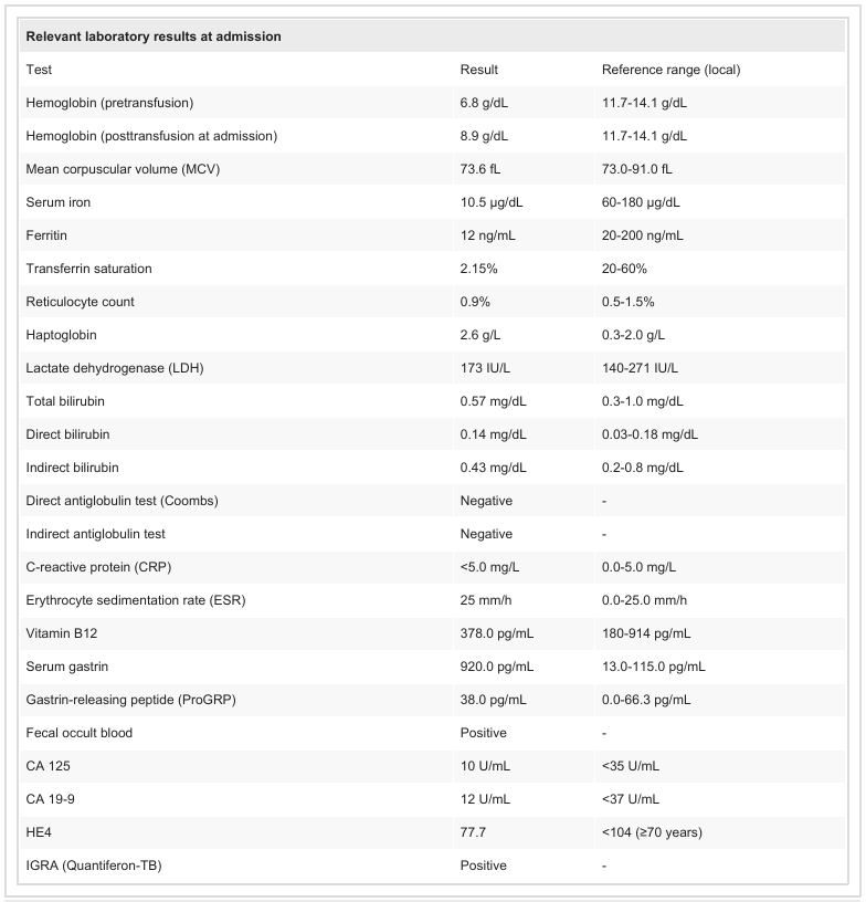
입원 시 시행한 검사에서 지속적인 소구성 빈혈과 함께 철 및 트랜스페린 포화도의 현저한 감소가 확인되어 철결핍성 빈혈(iron-deficiency anemia)에 합당한 소견을 보였다. 용혈(hemolysis)을 시사하는 검사 이상은 없었다. 간, 신장, 전해질 검사 결과는 모두 정상 범위였으며, 염증 표지자(inflammatory markers)는 상승하지 않았다. 비타민 B12 수치는 정상 범위로 유지되었다. 혈청 가스트린(serum gastrin)은 현저히 상승되어 있었으나, 가스트린 방출 펩타이드(gastrin-releasing peptide)는 정상 범위 내에 있었다(Table 1).
TABLE 1: Laboratory parameters at hospital admission CRP: C-reactive protein; ESR: erythrocyte sedimentation rate; LDH: lactate dehydrogenase; MCV: mean corpuscular volume; ProGRP: gastrin-releasing peptide; HE4: human epididymis protein 4; IGRA: interferon gamma release assay; CA 125: cancer antigen 125
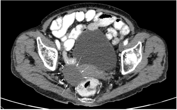
진단적 평가의 일환으로 시행한 복부-골반 전산화단층촬영(abdominopelvic computed tomography)에서 경계가 명확하고 국소 침윤 소견이 없는 우측 난소 낭종(right ovarian cyst)이 확인되었다(Figure 1). 암항원 125 (CA-125), CA 19-9, 그리고 암태아성항원(carcinoembryonic antigen, CEA)을 포함한 종양표지자(tumor markers)는 모두 정상 범위였으며, 부인과 팀에서 악성 종양 가능성은 배제되었다(Table 1).
FIGURE 1: Abdominopelvic CT showing right ovarian cyst
Well-defined cyst with regular borders and no local invasion
인터페론-감마 분비검사(interferon-gamma release assay, IGRA)에서 양성 소견이 확인되어 활동성 결핵 가능성을 평가하기 위해 흉부 X선 검사, 흉복부 전산화단층촬영(thoracoabdominopelvic CT), 경부 초음파(cervical ultrasound)를 시행하였으나 모두 정상 소견을 보였다. 유도 객담(induced sputum) 3회 검사에서도 항산균(acid-fast bacilli)은 검출되지 않았다.
상부위장관내시경(upper gastrointestinal endoscopy)에서는 위저부(fundus)와 체부(body)에서 위축성 점막(atrophic mucosa)이 관찰되었으며, 혈관 분포가 증가된 다수의 무경성(sessile) 용종이 확인되었다(Figures 2, 3). 이에 대해 생검이 시행되었다. 대장내시경(colonoscopy)에서는 대장 질환(colorectal pathology)이 배제되었다.
FIGURE 2: Endoscopic image of the gastric fundus showing a small, sessile, hypervascular polyp
The lesion was found during routine esophagogastroduodenoscopy in an elderly patient with anemia.
The surrounding mucosa shows signs of atrophy.
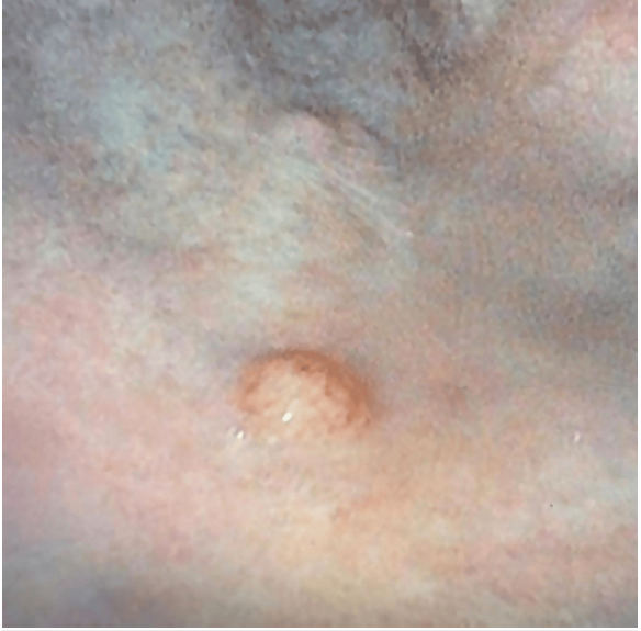
FIGURE 3: Closer endoscopic view of a hypervascular polyp in the gastric body
The smooth and well-circumscribed appearance is characteristic of type 1 gastric neuroendocrine tumors.

환자는 양성자펌프억제제(proton pump inhibitor, PPI)를 복용하고 있지 않아 약물 유발성 고가스트린혈증(drug-induced hypergastrinemia)의 가능성을 배제할 수 있었고, 자가면역성 위축성 위염(autoimmune atrophic gastritis)이 근본 원인일 가능성을 뒷받침하였다.
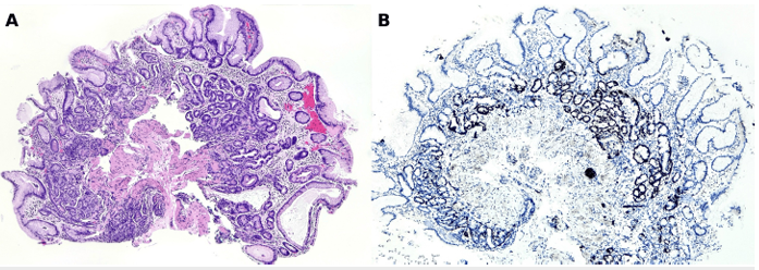
위 생검 조직의 조직학적 평가(histological evaluation)에서 위저부와 체부에 장형화생(intestinal metaplasia)과 유문선화생(pyloric metaplasia)을 동반한 만성 위축성 위염(chronic atrophic gastritis)이 확인되었다. 여러 부위에서 ECL 세포의 선상(linear) 및 국소 결절성(nodular) 과증식이 관찰되었으며(Figure 4A), 시냅토피신(synaptophysin)의 강한 발현이 확인되었다(Figure 4B).
FIGURE 4: Hyperplasia of gastric enterochromaffin-like (ECL) cells
(A) Gastric mucosa showing increased numbers of ECL cells distributed along the base of the fundic glands.
(B) Immunohistochemistry for synaptophysin showing strong and diffuse cytoplasmic expression in the hyperplastic neuroendocrine cells, with a linear and focally nodular pattern
다른 생검 조각들에서는 점막(mucosa)과 점막하층(submucosa)을 침윤하고, 일부 근육점막층(muscularis mucosae)까지 국소적으로 확장된 세포들이 고형(solid) 및 세주형(trabecular) 군집을 이루는 분화가 좋은 신경내분비종양(well-differentiated neuroendocrine tumor) 이 확인되었다(Figure 5A). 고배율 관찰(Figure 5B)에서 종양세포들은 둥근 핵(round nuclei), 미세립상 염색질(finely granular “salt and pepper” chromatin), 그리고 소량의 호산성 세포질(scant eosinophilic cytoplasm)을 보였다. 면역조직화학염색(immunohistochemistry)에서는 chromogranin(Figure 5C)과 synaptophysin이 양성으로 확인되었으며, Ki-67 증식지수(proliferation index) 는 5%로 측정되어 세계보건기구(WHO) 기준의 2등급(grade II) 신경내분비종양에 합당한 소견이었다. 특수염색(special stain)에서는 Helicobacter pylori가 검출되지 않았다.
FIGURE 5: Gastric neuroendocrine tumor in endoscopic biopsy
(A) Gastric mucosa showing a solid and trabecular proliferation of tumor cells infiltrating the mucosa and submucosa, with focal extension into the muscularis mucosae. (B) At higher magnification, the nests are composed of cells with round nuclei, finely granular “salt and pepper” chromatin (characteristic of neuroendocrine tumors), and scant eosinophilic cytoplasm. (C) Immunohistochemistry for chromogranin showing strong cytoplasmic expression in tumor cells. (D) Ki-67 immunostaining showing a proliferation index of approximately 5% 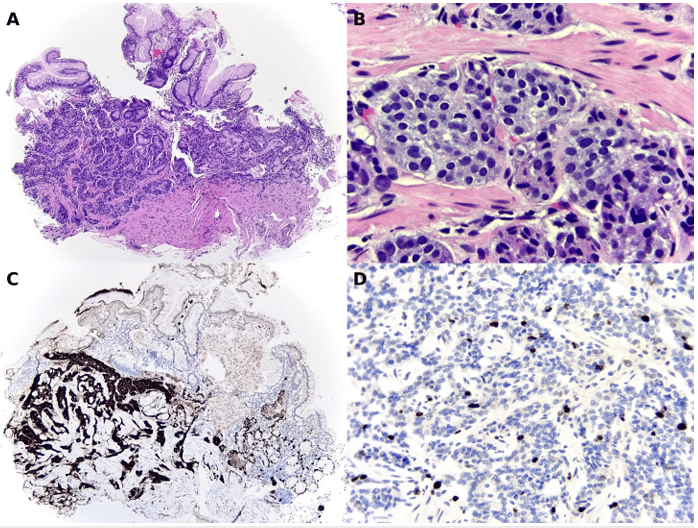
Discussion
제1형 위 신경내분비종양(Type 1 G-NET)은 전체 위 신경내분비종양의 약 80%를 차지하며, 자가면역성 만성 위축성 위염(autoimmune chronic atrophic gastritis) 과 밀접한 관련이 있다[2,3]. 그 병태생리학적 기전은 벽세포(parietal cell)의 점진적 소실, 저산증(hypochlorhydria), 그리고 만성 고가스트린혈증(chronic hypergastrinemia) 에 이차적으로 발생하는 ECL 세포 과증식(ECL cell hyperplasia) 으로 이어지며, 결국 신생물(neoplasia)로 발전하게 된다[1,3]. 이러한 병변은 대개 작고, 다발성이며, 위저부(fundus)와 체부(body)에 국한되어 있고, 전이 가능성(metastatic potential) 은 낮은 편이다[5].
정확한 임상적 및 조직학적 분류가 매우 중요하다. 세계보건기구(WHO) 는 임상 아형에 관계없이 Ki-67 증식지수와 유사분열수(mitotic count) 를 기준으로 종양을 1, 2, 3등급으로 분류할 것을 권고하고 있다[4]. 진단은 가스트린(gastrin) 과 가스트린 방출 펩타이드(gastrin-releasing peptide, GRP) 수치, 그리고 자가면역적 배경을 간접적으로 반영하는 비타민 B12 수치 평가를 통해 보조적으로 뒷받침된다[3,6]. 제1형 G-NET과 가장 흔히 연관되는 임상적 상태는 만성 자가면역성 위축성 위염이며, 본 증례에서도 동일하게 확인되었다.
이 증례는 비특이적 증상과 심한 철결핍성 빈혈(iron-deficiency anemia) 을 보인 허약한 고령 환자(frail elderly patient) 의 사례를 보여준다. 감염성 질환, 대장 및 부인과 악성종양을 배제한 뒤, 내시경 및 조직학적 소견과 가스트린 수치의 상승을 종합하여 자가면역성 위염과 연관된 제1형 G-NET으로 진단되었다.
치료는 종양의 크기(size), 등급(grade), 재발 여부(recurrence) 에 따라 개별적으로 결정된다. 대부분의 작고 저등급(low-grade) 제1형 G-NET은 내시경 추적관찰(endoscopic surveillance) 또는 국소 절제(local resection) 로 관리가 가능하다[5]. 반면, 크거나 재발성 병변의 경우에는 전정부절제술(antrectomy) 또는 위부분절제술(subtotal gastrectomy) 이 필요할 수 있다[8].
특히 고령 또는 허약한 환자에서는 치료의 위험과 이득을 신중히 평가하기 위해 기능적 상태(functional status) 와 기대수명(life expectancy) 에 대한 평가가 중요하다. 기존 문헌에서도 이러한 경우에 개별화된 접근(individualized approach) 의 필요성이 강조되고 있다[9,10]. 본 증례에서는 병변의 저등급, 작은 크기, 전이 부재 등을 고려하여, 다학제팀(multidisciplinary team) 이 내시경 추적관찰을 가장 적절한 관리 전략으로 선택하였다.
Conclusions
이 증례는 고령 환자에서 비특이적 위장관 증상(nonspecific gastrointestinal symptoms) 과 심한 철결핍성 빈혈(severe iron-deficiency anemia) 이 동반된 경우, 위 신경내분비종양(G-NET) 을 감별진단(differential diagnosis) 에 포함시킬 필요성을 강조한다.
비록 제1형 G-NET(Type 1 G-NET) 은 일반적으로 저등도(indolent) 의 임상 경과를 보이지만, 정확한 진단을 위해서는 내시경(endoscopy), 조직학적 평가(histology), 그리고 생화학적 표지자(biochemical markers) 를 포함한 체계적인 진단 접근(systematic diagnostic approach) 이 필수적이다.
치료 전략은 종양의 특성(tumor characteristics), 재발 위험(recurrence risk), 그리고 특히 환자의 기능적 상태(functional status) 와 동반 질환(comorbidities) 을 고려하여 개별화되어야 한다.
이러한 환자군에서 안전하고 적절한 치료 결정을 보장하기 위해 다학제적 평가(multidisciplinary evaluation) 가 필수적이다. 본 증례는 노인종양학(geriatric oncology) 분야에서 개별화된 진료(individualized care) 의 중요성을 다시 한 번 부각시킨다.
<Abstract>
Gastric neuroendocrine tumors (G-NETs) are rare neoplasms with increasing incidence due to the broader use of endoscopy and improved diagnostic sensitivity. Type 1 G-NETs are the most common subtype and are typically associated with chronic atrophic autoimmune gastritis and hypergastrinemia. We present the case of an 84-year-old female patient admitted for the evaluation of progressive fatigue, weight loss, anorexia, and intermittent diarrhea. Initial workup revealed severe iron-deficiency anemia. Further testing demonstrated a positive interferon-gamma release assay (IGRA), with no evidence of active tuberculosis. Colonoscopy and gynecologic evaluation ruled out colorectal and adnexal malignancies. Esophagogastroduodenoscopy identified multiple raised vascular lesions in the gastric fundus and body. Histopathology confirmed a well-differentiated type 1 G-NET (grade 2, Ki-67 index 5%), associated with enterochromaffin-like cell hyperplasia and autoimmune atrophic gastritis. The patient was managed through multidisciplinary evaluation. Given the small size, low grade, and absence of metastasis, a conservative approach with endoscopic surveillance was chosen. While endoscopic resection or surgical interventions such as antrectomy or gastrectomy may be appropriate in selected cases, functional assessment and individualized decision-making remain essential, particularly in older adults with multimorbidity. This case highlights the diagnostic complexity of type 1 G-NETs in elderly patients and underscores the importance of tailored, multidisciplinary management strategies.
Keywords: anemia; atrophic gastritis; elderly; gastric neuroendocrine tumor; hypergastrinemia.


Cortés-Marín EE, González-Rodríguez JC, Cristofori M, Antunez Oliva JA, Vargas Wille MF.
Cureus. 2025 Jul 7;17(7):e87429.
Introduction
위장 신경내분비종양(Gastric neuroendocrine tumors, G-NETs)은 드물게 발생하는 임상병리학적 질환으로, 최근 수십 년간 내시경의 보편화와 진단 인식의 향상으로 인해 그 발생률이 증가하고 있다. G-NETs는 전체 위 신생물의 약 1~2%를 차지하며, 위저부(fundus)와 체부(corpus)에 위치한 엔테로크로마핀 유사(enterochromaffin-like, ECL) 세포에서 기원한다[1].
임상적 및 병태생리학적으로 G-NETs는 세 가지 주요 아형으로 분류된다. 제1형(type 1)은 전체의 70~80%를 차지하며, 만성 자가면역성 위축성 위염(chronic autoimmune atrophic gastritis), 고가스트린혈증(hypergastrinemia), 그리고 ECL 세포 과증식(ECL cell hyperplasia)과 연관되어 있다[2]. 제2형(type 2)은 다발성 내분비종양증 1형(MEN-1)과 연관된 졸링거-엘리슨 증후군(Zollinger-Ellison syndrome)과 관련이 있으며, 제3형(type 3)은 산발적으로 발생하는 가스트린 비의존성 종양으로 보다 공격적인 생물학적 행동과 높은 전이 가능성을 보인다[3].
이러한 종양은 아형, 조직학적 등급, 종양 크기, 전이 여부 등에 따라 치료 전략이 크게 달라지기 때문에 정확한 인지가 필수적이다[4,5]. 실제 임상에서는 고령 환자나 복합질환을 가진 환자에서 비특이적인 위장관 증상으로 발현되거나 우연히 발견되는 경우가 있어 진단이 특히 어려울 수 있다[6,7]. 본 보고에서는 자가면역성 위염의 배경에서 제1형 G-NET으로 진단된 고령 여성의 증례를 제시하며, 고령 환자에서의 개별화된 진단 및 치료 접근의 중요성을 강조하고자 한다.
Case presentation
84세 여성 환자가 전신성 고혈압 병력을 가지고 있었으며, 약 8개월 동안 점진적인 피로, 비의도적 체중 감소, 식욕 부진, 간헐적 설사를 호소하였다. 암, 결핵, 자가면역질환 또는 자가염증질환의 가족력은 없었다. 신체검사에서 영양 상태가 불량해 보였다. 초기 검사에서 대변잠혈검사(fecal occult blood test)가 양성이었으며, 현저한 소구성 저색소성 빈혈(microcytic hypochromic anemia)이 확인되어 농축적혈구(packed red blood cells) 2단위의 수혈이 필요하였다. 환자는 추가 평가를 위해 입원하였다.
입원 시 시행한 검사에서 지속적인 소구성 빈혈과 함께 철 및 트랜스페린 포화도의 현저한 감소가 확인되어 철결핍성 빈혈(iron-deficiency anemia)에 합당한 소견을 보였다. 용혈(hemolysis)을 시사하는 검사 이상은 없었다. 간, 신장, 전해질 검사 결과는 모두 정상 범위였으며, 염증 표지자(inflammatory markers)는 상승하지 않았다. 비타민 B12 수치는 정상 범위로 유지되었다. 혈청 가스트린(serum gastrin)은 현저히 상승되어 있었으나, 가스트린 방출 펩타이드(gastrin-releasing peptide)는 정상 범위 내에 있었다(Table 1).
TABLE 1: Laboratory parameters at hospital admission CRP: C-reactive protein; ESR: erythrocyte sedimentation rate; LDH: lactate dehydrogenase; MCV: mean corpuscular volume; ProGRP: gastrin-releasing peptide; HE4: human epididymis protein 4; IGRA: interferon gamma release assay; CA 125: cancer antigen 125
진단적 평가의 일환으로 시행한 복부-골반 전산화단층촬영(abdominopelvic computed tomography)에서 경계가 명확하고 국소 침윤 소견이 없는 우측 난소 낭종(right ovarian cyst)이 확인되었다(Figure 1). 암항원 125 (CA-125), CA 19-9, 그리고 암태아성항원(carcinoembryonic antigen, CEA)을 포함한 종양표지자(tumor markers)는 모두 정상 범위였으며, 부인과 팀에서 악성 종양 가능성은 배제되었다(Table 1).
FIGURE 1: Abdominopelvic CT showing right ovarian cyst
Well-defined cyst with regular borders and no local invasion
인터페론-감마 분비검사(interferon-gamma release assay, IGRA)에서 양성 소견이 확인되어 활동성 결핵 가능성을 평가하기 위해 흉부 X선 검사, 흉복부 전산화단층촬영(thoracoabdominopelvic CT), 경부 초음파(cervical ultrasound)를 시행하였으나 모두 정상 소견을 보였다. 유도 객담(induced sputum) 3회 검사에서도 항산균(acid-fast bacilli)은 검출되지 않았다.
상부위장관내시경(upper gastrointestinal endoscopy)에서는 위저부(fundus)와 체부(body)에서 위축성 점막(atrophic mucosa)이 관찰되었으며, 혈관 분포가 증가된 다수의 무경성(sessile) 용종이 확인되었다(Figures 2, 3). 이에 대해 생검이 시행되었다. 대장내시경(colonoscopy)에서는 대장 질환(colorectal pathology)이 배제되었다.
FIGURE 2: Endoscopic image of the gastric fundus showing a small, sessile, hypervascular polyp
The lesion was found during routine esophagogastroduodenoscopy in an elderly patient with anemia.
The surrounding mucosa shows signs of atrophy.
FIGURE 3: Closer endoscopic view of a hypervascular polyp in the gastric body
The smooth and well-circumscribed appearance is characteristic of type 1 gastric neuroendocrine tumors.
환자는 양성자펌프억제제(proton pump inhibitor, PPI)를 복용하고 있지 않아 약물 유발성 고가스트린혈증(drug-induced hypergastrinemia)의 가능성을 배제할 수 있었고, 자가면역성 위축성 위염(autoimmune atrophic gastritis)이 근본 원인일 가능성을 뒷받침하였다.
위 생검 조직의 조직학적 평가(histological evaluation)에서 위저부와 체부에 장형화생(intestinal metaplasia)과 유문선화생(pyloric metaplasia)을 동반한 만성 위축성 위염(chronic atrophic gastritis)이 확인되었다. 여러 부위에서 ECL 세포의 선상(linear) 및 국소 결절성(nodular) 과증식이 관찰되었으며(Figure 4A), 시냅토피신(synaptophysin)의 강한 발현이 확인되었다(Figure 4B).
FIGURE 4: Hyperplasia of gastric enterochromaffin-like (ECL) cells
(A) Gastric mucosa showing increased numbers of ECL cells distributed along the base of the fundic glands.
(B) Immunohistochemistry for synaptophysin showing strong and diffuse cytoplasmic expression in the hyperplastic neuroendocrine cells, with a linear and focally nodular pattern
다른 생검 조각들에서는 점막(mucosa)과 점막하층(submucosa)을 침윤하고, 일부 근육점막층(muscularis mucosae)까지 국소적으로 확장된 세포들이 고형(solid) 및 세주형(trabecular) 군집을 이루는 분화가 좋은 신경내분비종양(well-differentiated neuroendocrine tumor) 이 확인되었다(Figure 5A). 고배율 관찰(Figure 5B)에서 종양세포들은 둥근 핵(round nuclei), 미세립상 염색질(finely granular “salt and pepper” chromatin), 그리고 소량의 호산성 세포질(scant eosinophilic cytoplasm)을 보였다. 면역조직화학염색(immunohistochemistry)에서는 chromogranin(Figure 5C)과 synaptophysin이 양성으로 확인되었으며, Ki-67 증식지수(proliferation index) 는 5%로 측정되어 세계보건기구(WHO) 기준의 2등급(grade II) 신경내분비종양에 합당한 소견이었다. 특수염색(special stain)에서는 Helicobacter pylori가 검출되지 않았다.
FIGURE 5: Gastric neuroendocrine tumor in endoscopic biopsy
(A) Gastric mucosa showing a solid and trabecular proliferation of tumor cells infiltrating the mucosa and submucosa, with focal extension into the muscularis mucosae. (B) At higher magnification, the nests are composed of cells with round nuclei, finely granular “salt and pepper” chromatin (characteristic of neuroendocrine tumors), and scant eosinophilic cytoplasm. (C) Immunohistochemistry for chromogranin showing strong cytoplasmic expression in tumor cells. (D) Ki-67 immunostaining showing a proliferation index of approximately 5%
Discussion
제1형 위 신경내분비종양(Type 1 G-NET)은 전체 위 신경내분비종양의 약 80%를 차지하며, 자가면역성 만성 위축성 위염(autoimmune chronic atrophic gastritis) 과 밀접한 관련이 있다[2,3]. 그 병태생리학적 기전은 벽세포(parietal cell)의 점진적 소실, 저산증(hypochlorhydria), 그리고 만성 고가스트린혈증(chronic hypergastrinemia) 에 이차적으로 발생하는 ECL 세포 과증식(ECL cell hyperplasia) 으로 이어지며, 결국 신생물(neoplasia)로 발전하게 된다[1,3]. 이러한 병변은 대개 작고, 다발성이며, 위저부(fundus)와 체부(body)에 국한되어 있고, 전이 가능성(metastatic potential) 은 낮은 편이다[5].
정확한 임상적 및 조직학적 분류가 매우 중요하다. 세계보건기구(WHO) 는 임상 아형에 관계없이 Ki-67 증식지수와 유사분열수(mitotic count) 를 기준으로 종양을 1, 2, 3등급으로 분류할 것을 권고하고 있다[4]. 진단은 가스트린(gastrin) 과 가스트린 방출 펩타이드(gastrin-releasing peptide, GRP) 수치, 그리고 자가면역적 배경을 간접적으로 반영하는 비타민 B12 수치 평가를 통해 보조적으로 뒷받침된다[3,6]. 제1형 G-NET과 가장 흔히 연관되는 임상적 상태는 만성 자가면역성 위축성 위염이며, 본 증례에서도 동일하게 확인되었다.
이 증례는 비특이적 증상과 심한 철결핍성 빈혈(iron-deficiency anemia) 을 보인 허약한 고령 환자(frail elderly patient) 의 사례를 보여준다. 감염성 질환, 대장 및 부인과 악성종양을 배제한 뒤, 내시경 및 조직학적 소견과 가스트린 수치의 상승을 종합하여 자가면역성 위염과 연관된 제1형 G-NET으로 진단되었다.
치료는 종양의 크기(size), 등급(grade), 재발 여부(recurrence) 에 따라 개별적으로 결정된다. 대부분의 작고 저등급(low-grade) 제1형 G-NET은 내시경 추적관찰(endoscopic surveillance) 또는 국소 절제(local resection) 로 관리가 가능하다[5]. 반면, 크거나 재발성 병변의 경우에는 전정부절제술(antrectomy) 또는 위부분절제술(subtotal gastrectomy) 이 필요할 수 있다[8].
특히 고령 또는 허약한 환자에서는 치료의 위험과 이득을 신중히 평가하기 위해 기능적 상태(functional status) 와 기대수명(life expectancy) 에 대한 평가가 중요하다. 기존 문헌에서도 이러한 경우에 개별화된 접근(individualized approach) 의 필요성이 강조되고 있다[9,10]. 본 증례에서는 병변의 저등급, 작은 크기, 전이 부재 등을 고려하여, 다학제팀(multidisciplinary team) 이 내시경 추적관찰을 가장 적절한 관리 전략으로 선택하였다.
Conclusions
이 증례는 고령 환자에서 비특이적 위장관 증상(nonspecific gastrointestinal symptoms) 과 심한 철결핍성 빈혈(severe iron-deficiency anemia) 이 동반된 경우, 위 신경내분비종양(G-NET) 을 감별진단(differential diagnosis) 에 포함시킬 필요성을 강조한다.
비록 제1형 G-NET(Type 1 G-NET) 은 일반적으로 저등도(indolent) 의 임상 경과를 보이지만, 정확한 진단을 위해서는 내시경(endoscopy), 조직학적 평가(histology), 그리고 생화학적 표지자(biochemical markers) 를 포함한 체계적인 진단 접근(systematic diagnostic approach) 이 필수적이다.
치료 전략은 종양의 특성(tumor characteristics), 재발 위험(recurrence risk), 그리고 특히 환자의 기능적 상태(functional status) 와 동반 질환(comorbidities) 을 고려하여 개별화되어야 한다.
이러한 환자군에서 안전하고 적절한 치료 결정을 보장하기 위해 다학제적 평가(multidisciplinary evaluation) 가 필수적이다. 본 증례는 노인종양학(geriatric oncology) 분야에서 개별화된 진료(individualized care) 의 중요성을 다시 한 번 부각시킨다.
<Abstract>
Gastric neuroendocrine tumors (G-NETs) are rare neoplasms with increasing incidence due to the broader use of endoscopy and improved diagnostic sensitivity. Type 1 G-NETs are the most common subtype and are typically associated with chronic atrophic autoimmune gastritis and hypergastrinemia. We present the case of an 84-year-old female patient admitted for the evaluation of progressive fatigue, weight loss, anorexia, and intermittent diarrhea. Initial workup revealed severe iron-deficiency anemia. Further testing demonstrated a positive interferon-gamma release assay (IGRA), with no evidence of active tuberculosis. Colonoscopy and gynecologic evaluation ruled out colorectal and adnexal malignancies. Esophagogastroduodenoscopy identified multiple raised vascular lesions in the gastric fundus and body. Histopathology confirmed a well-differentiated type 1 G-NET (grade 2, Ki-67 index 5%), associated with enterochromaffin-like cell hyperplasia and autoimmune atrophic gastritis. The patient was managed through multidisciplinary evaluation. Given the small size, low grade, and absence of metastasis, a conservative approach with endoscopic surveillance was chosen. While endoscopic resection or surgical interventions such as antrectomy or gastrectomy may be appropriate in selected cases, functional assessment and individualized decision-making remain essential, particularly in older adults with multimorbidity. This case highlights the diagnostic complexity of type 1 G-NETs in elderly patients and underscores the importance of tailored, multidisciplinary management strategies.
Keywords: anemia; atrophic gastritis; elderly; gastric neuroendocrine tumor; hypergastrinemia.